My PhD Research
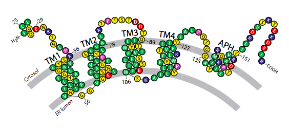
Tubule formation in the ER was shown by Voeltz et al (2006) to require a class of membrane proteins with unusually short transmembrane (TM) regions. These short TMs are thought to both “wedge” and “scaffold” ER membranes into their tubular architecture. My work demonstrated that a third component, amphipathic helices, is required for tubule formation and that this component is likely to be evolutionarily conserved.
The importance of ER tubules
The tubular ER comprises a highly dynamic, polygonal network of membrane tubules with roles in cell transport, signalling and metabolism. The ratio of sheet ER to tubular ER is cell type dependent. Cells specialised in metabolism such as hepatocytes contain mainly tubules whereas secretory cells (such as pancreatic acinar) contain primarily sheet ER. The composition of the ER can change dramatically during the cell cycle. For example during transition from interphase into mitosis the ER transforms from being primarily tubular to sheet!
ER tubules and disease
Dysfunction of the tubular ER network has been implicated in human diseases such as hereditary spastic paraplegia (HSP) and schizophrenia. In particular proteins involved in the maintanence and dynamics of ER tubules such as Spastin, Atlastin, REEPs and Reticulons have all been implicated in HSP.